How To Measure Haller Index At Home
- Technical Innovation
- Open Access
- Published:
MRI for the evaluation of pectus excavatum
Pediatric Radiology book 41,pages 757–758 (2011)Cite this article
Abstract
Pectus excavatum, the most common congenital deformity of the inductive chest wall, is both a cosmetic and functional abnormality. The degree of abnormal chest wall deformity determines its functional effect, particularly its cardiac and pulmonary touch on. Although CT scanning is the most widely used cross-sectional imaging technique used to measure out the Haller alphabetize, the radiation exposure is reason to seek other alternatives. At our institution, we take introduced a rapid MRI technique for this purpose, which utilizes a unmarried-axial 2-D FIESTA acquisition.
Introduction
Pectus excavatum, the nearly common built deformity of the anterior chest wall, is both a cosmetic and functional abnormality. The degree of aberrant chest wall deformity determines its functional issue, specially its cardiac and pulmonary impact.
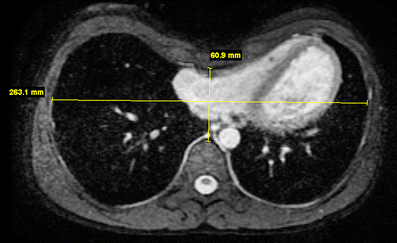
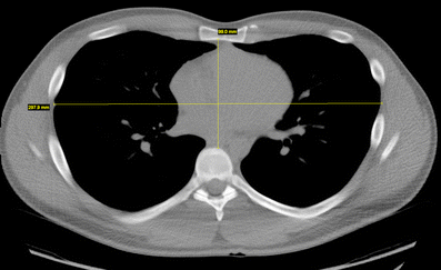
The diagnosis of pectus excavatum is clinical. Withal, the quantitative measurement of the deformity is measured using cross-sectional imaging. In particular, the ordinarily used Haller index is the ratio betwixt the lateral distance of the chest wall (inner margins) and the narrowest anteroposterior distance between the vertebrae and sternum (both measured at the same centric level). If this ratio is above 3.25, it is considered astringent [1].
CT scanning has commonly been used for this purpose [i]. Contempo reports in the literature, still, have recognized the problem of radiation exposure when using CT scanning for this purpose [two].
Description
At our institution, we have introduced the use of a fast MRI technique to measure the Haller index. This technique has proven reliable and like shooting fish in a barrel to perform, and avoids the ionizing radiations of CT scanning.
MRI scanning at our institution for this purpose is performed on a 1.5 T HDxT platform (General Electric, Milwaukee, WI). An 8- or 12-channel trunk scroll is used. A brusque localizer (ten s) is followed by a two-D FIESTA (TE 1.7–1.8, TR 3.7–3.8, 192 x 256, 1 NEX) fat-saturated sequence. Slice thickness is five mm with a i mm gap obtained through the lower chest. The time required for the FIESTA sequence is approximately xx s for a 20-slice acquisition. Jiff-agree is not necessary within these scan parameters, although both inspiratory equally well as expiratory acquisitions could be easily obtained with a minimum of added effort. Cardiac gating is non needed. Full scan time is under 5 min. The specific MR sequence (FIESTA = Fast Imaging Employing Steady-state Conquering) was selected because it tin produce loftier SNR images in a curt acquisition time.
Measurement of the Haller alphabetize is performed in the standard style using the electronic calipers on our PACS monitors.
Word
At our institution, MRI has replaced CT scanning for evaluating pectus excavatum and the measurement of the Haller index. A curt localizer is followed by a single FIESTA (Phillips refers to this equally balanced FFE; Siemens refers to this as trueFISP) acquisition. The unabridged MRI examination requires less than 5 min to perform, which compares favorably with CT scanning. While both MRI and CT produce diagnostic quality scans which can exist easily interpreted, MRI requires no ionizing radiations. Since pectus excavatum repair is almost exclusively performed in older children and adolescents, no sedation is required.
At our site, our primary pectus excavatum surgeon has switched to ordering the MRI examination described higher up, and has reported that the results are reliable and describe well the relevant anatomy. To date, we have scanned approximately fifty patients using this MRI technique.
Other researchers have recently proposed using a two-view chest radiograph to replace CT scanning for the purpose of Haller index measurement. Nevertheless, this still requires the utilise of ionizing radiation [3].
Though in that location is an association of bronchiectasis with pectus excavatum [4], the primary purpose of presurgical imaging in this status is to appraise the Haller index. In fact, single-slice CT scanning would as well likely underestimate the degree of bronchiectasis. In patients with suspected bronchiectasis related to pectus excavatum, full-chest CT scanning may be the preferred presurgical prototype exam.
Future directions in the imaging evaluation of pectus excavatum include the routine acquisition of inspiratory and expiratory MRI sequences. Inquiry has shown that this may provide more physiological data; in expiration, the deformity may worsen [5]. Furthermore, cine MR imaging has been shown to be capable of evaluating both breast morphology and chest wall kinetics, and may well add of import diagnostic information [half dozen] (Figs 1 and 2).

Centric 2-D FIESTA of the chest demonstrates pectus excavatum with the Haller index measurement

Axial CT scan of the chest demonstrates pectus excavatum with the Haller index measurement
References
-
Haller JA Jr, Kramer SS, Lietman SA et al (1987) Utilise of CT scans in selection of patients for pectus excavatum surgery: a preliminary written report. J Pediatr Surg 22:904–906
-
Rattan AS, Laor T, Ryckman FC et al (2010) Pectus excavatum imaging: enough but not too much. Pediatr Radiol 40:168–172
-
Khanna Grand, Jaju A, Don S et al (2010) Comparison of Haller alphabetize values calculated with chest radiographs versus CT for pectus excavatum evaluation. Pediatr Radiol 40:1763–1767
-
Morshuis WJ, Mulder H, Wapperom G et al (1992) Pectus excavatum. A clinical written report with long-term postoperative follow-up. Eur J Cardiothorac Surg 6:318–328, discussion 328–329
-
Raichura Due north, Entwisle J, Leverment J et al (2001) Breath-hold MRI in evaluating patients with pectus excavatum. Br J Radiol 74:701–708
-
Herrmann KA, Zech C, Strauss T et al (2006) Cine MRI of the thorax in patients with pectus excavatum. Radiologe 46:309–316, German
Open Access
This article is distributed under the terms of the Creative Commons Attribution Noncommercial License which permits whatsoever noncommercial use, distribution, and reproduction in whatever medium, provided the original author(southward) and source are credited.
Author information
Authors and Affiliations
Corresponding author
Rights and permissions
Open Access This is an open access article distributed under the terms of the Creative Eatables Attribution Noncommercial License (https://creativecommons.org/licenses/by-nc/2.0), which permits any noncommercial use, distribution, and reproduction in any medium, provided the original writer(s) and source are credited.
Reprints and Permissions
Most this article
Cite this article
Marcovici, P.A., LoSasso, B.Due east., Kruk, P. et al. MRI for the evaluation of pectus excavatum. Pediatr Radiol 41, 757–758 (2011). https://doi.org/10.1007/s00247-011-2031-5
-
Received:
-
Revised:
-
Accustomed:
-
Published:
-
Issue Date:
-
DOI : https://doi.org/10.1007/s00247-011-2031-5
Keywords
- Pectus excavatum
- MRI
- Haller index
Source: https://link.springer.com/article/10.1007/s00247-011-2031-5

0 Response to "How To Measure Haller Index At Home"
Post a Comment